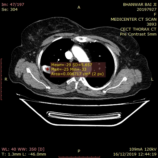
Imaging features :
There is well defined lobulated solitary soft tissue lesion of size 17 x 35 x 26 mm in the right upper lobe. There are multiple areas of popcorn like calcification. Few areas also show fat attenuation(Upto-20 HU). The lesion shows mild enhancement on post contrast study. The lesion is extending upto the periphery of the lung and abutting the major fissure.
Few centrilobular nodules with ground glass opacities in right middle lobe.
The small area of fibro parenchymal thickening in the lingular segment of left upper lobe, suggest sequelae of old broncho-pulmonary infection.
Rest of the lung appears normal with even distribution of the pulmonary arterial branches and that of the bronchial tree.
The anatomical configuration of the structures in the mediastinum and both hilar regions are normal.
There are subcentimeteric enlarged lymph nodes in pretracheal, right paratracheal & subcarinal region -?Reactive.
The arch of the aorta and the vessels arising from it are normal.
There is no evidence of pleural effusion seen.
Visualized abdomen shows no significant abnormality.
Bilateral adrenal glands are normal.
Visualized bones shows degenerative changes with diffuse idiopathic skeletal hyperostosis.
IMPRESSION: Above described CECT imaging findings are suggestive of
- Pulmonary hamartoma in right upper lobe.
- Few centrilobular nodules with ground glass opacities in right middle lobe, suggestive of infective etiology/bronchitis.
- Fibro parenchymal thickening in the lingular segment of left upper lobe, suggest sequelae of old broncho-pulmonary infection.

14 yr old female with swelling breast Fnac is suggestive of metastatic acute lymphoblastic leukemia ( rare tumor)
Read More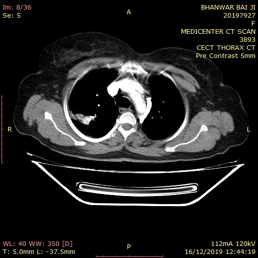
C.T.SCANImaging Features :
There is thick walled cavitatory lesion with spiculated borders in the apical segment of right upper lobe, measuring 50 x 48 x 51 mm. The lesion is causing stranding posterosuperiorly with infiltration & erosion of transverse process of D2, costovertebral joint and adjacent posterior 2nd rib & also erosion of the adjacent 3rd rib. Findings are suggestive of apical lung tumor (Pancost tumor). (Adv : HPE correlation)
There are large fibrocavitatory parenchymal thickening with dystrophic calcification, bronchiectasis, subpleural consolidation with adjacent architectural distortion seen in the left upper lobe. Multiple centrilobular nodules few showing tree in bud pattern & calcification, bronchiectasis, peribronchial thickening, and interlobular interstitial thickening in both lungs. Finding are suggestive of post primary tuberculosis.
There is well defined oval shaped soft tissue density lesion of size 9 x 9 mm seen in the dependent part of the left upper lobe cavity, suggestive of aspergilloma .
Trachea mildly shifted towards right side
Centriacenar emphysematous changes are seen in both lungs.
There are few enlarged lymph nodes noted involving prevascular, pretracheal, bilateral paratracheal, AP window, precarinal & subcarinal regions, largest measures 15 x 10 mm in the subcarinal region, ? Granulomatous ? Metastatic
The arch of the aorta and the vessels arising from it are normal.There is no evidence of pleural effusion seen.
Note is made of sliding hiatus hernia.
Visualized abdomen shows bilateral simple renal cortical cysts.
Visualized bones show old united fracture in the left 11th & 12th ribs posteriorly.
Impression: Above described CT imaging features reveal
- Right apical lung tumor (Pancost tumor) as described above. (Adv : HPE correlation)
- Post primary tuberculosis with aspergilloma in left upper lobe cavity.
- Diffuse centriacenar emphysematous changes.
- Mediastinal lymphadenopathy ? Granulomatous ? Metastatic
- Sliding hiatus hernia.
- Bilateral simple renal cortical cysts.
- Old united fracture in the left 11th & 12th ribs posteriorly.
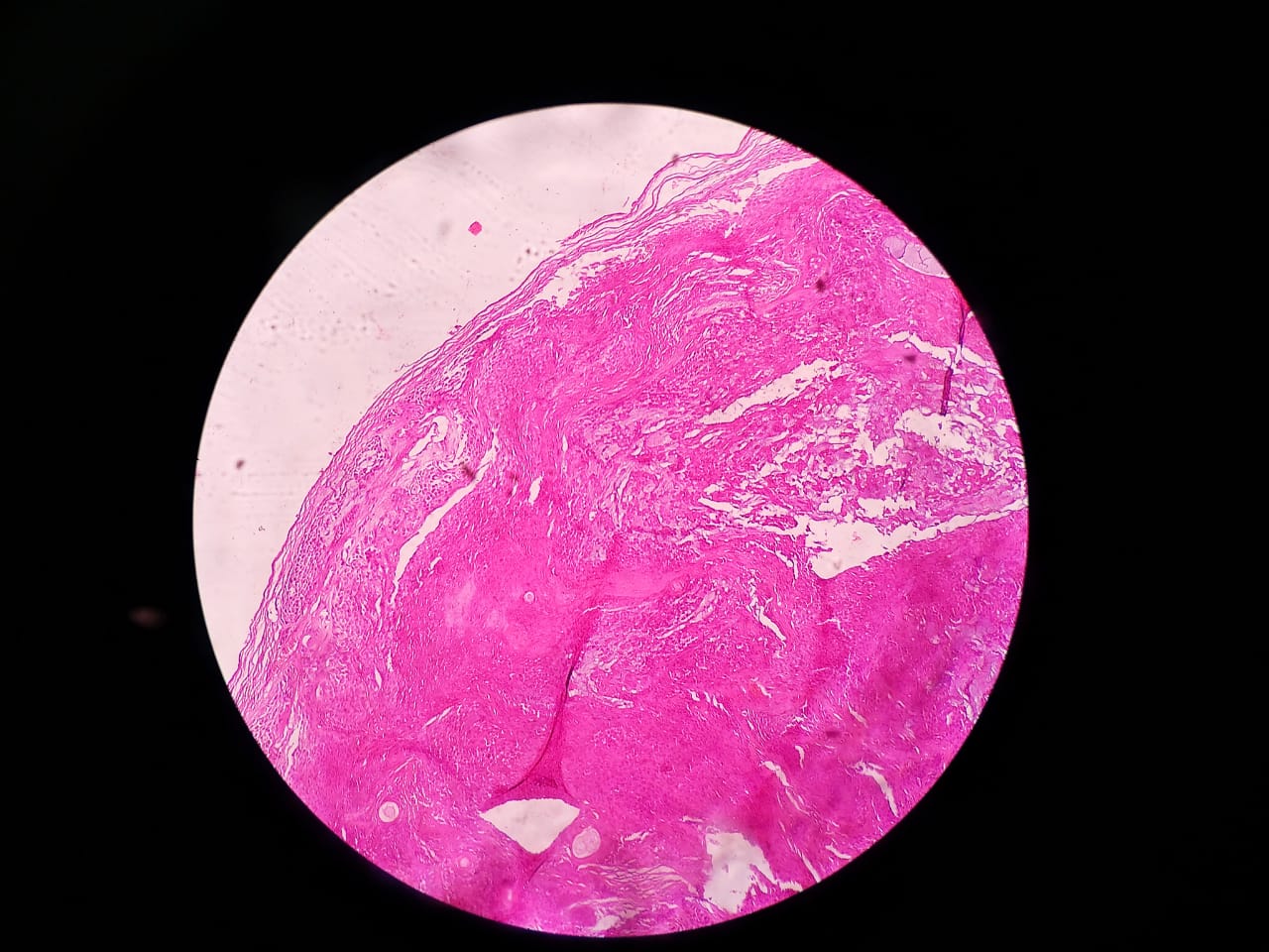
60 year male presented with slow growing swelling over left ear ( pinna).Histopathology was suggestive of Chondroid Syringoma.It is a benign tumor of sweat glands Ear is an unusual site for this tumor.
Read More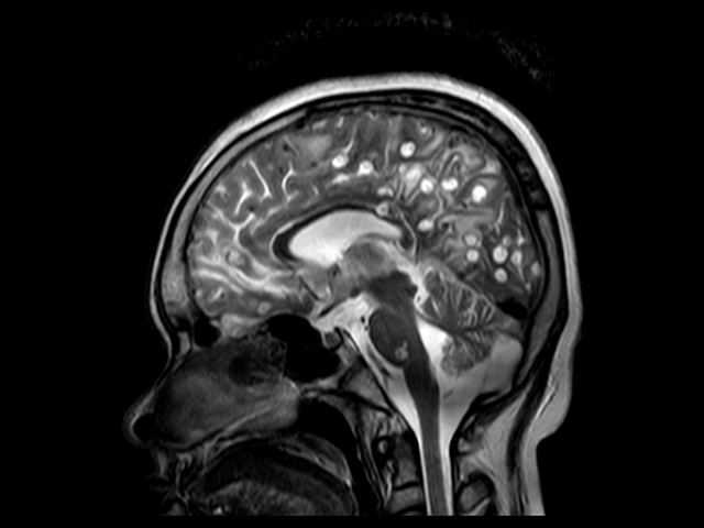
IMAGING FEATURE:
The study shows innumerable well circumscribed subcentimeteric cystic lesions diffusely studded in brain parenchyma involving all the lobes, capsuloganglionic region, right thalamus, corpus callosum, pons and cerebellum with significant perifocal and white matter edema. The lesions appear hyperintense on T2W with central T1W hyperintense signal corresponding gradient susceptibility on FFE, suggestive of calcification. On post contrast study there is thick enhancement of the wall with tiny central enhancing dot
No abnormal leptomeningeal enhancement is seen.
Rest of the brain parenchyma appears normal.
Ventricular drainage tube is noted on right side with its tip seen at the foramen of monro and traversing through the right lateral ventricle and frontal lobe into the frontal scalp region. There is mild dilatation of left lateral ventricle with midline shift of 8.0 mm towards right side. Right lateral ventricle is partially effaced. Also there is mild prominent of 3rd & 4th ventricle.
Conclusion:-
Above MR imaging findings suggestive of infective granuloma likely neurocysticercosis at colloid vesicular & granular nodular stage.
Read More
.jpeg)
Leptomeningeal myelomatosis: A rare case
A Middle aged male presented with altered sensorium without any signs of renal function deterioration
Csf examination showed monotonous population of malignant plasma cells.
MEDICENTRE SONOGRAPHY AND CLINICAL LAB
UDAIPUR RAJASTHAN
Read More