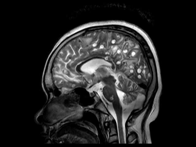
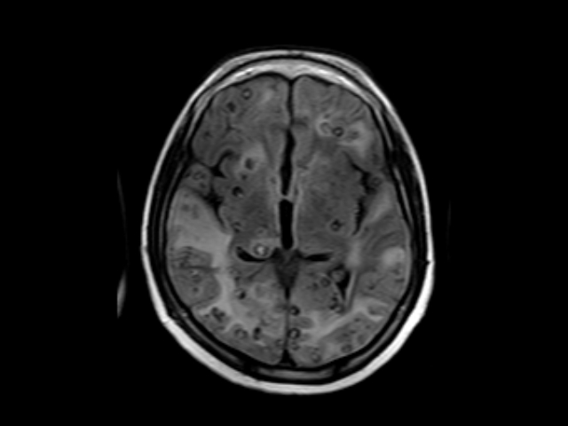
IMAGING FEATURE:
The study shows innumerable well circumscribed subcentimeteric cystic lesions diffusely studded in brain parenchyma involving all the lobes, capsuloganglionic region, right thalamus, corpus callosum, pons and cerebellum with significant perifocal and white matter edema. The lesions appear hyperintense on T2W with central T1W hyperintense signal corresponding gradient susceptibility on FFE, suggestive of calcification. On post contrast study there is thick enhancement of the wall with tiny central enhancing dot
No abnormal leptomeningeal enhancement is seen.
Rest of the brain parenchyma appears normal.
Ventricular drainage tube is noted on right side with its tip seen at the foramen of monro and traversing through the right lateral ventricle and frontal lobe into the frontal scalp region. There is mild dilatation of left lateral ventricle with midline shift of 8.0 mm towards right side. Right lateral ventricle is partially effaced. Also there is mild prominent of 3rd & 4th ventricle.
Conclusion:-
Above MR imaging findings suggestive of infective granuloma likely neurocysticercosis at colloid vesicular & granular nodular stage.