Imaging features :
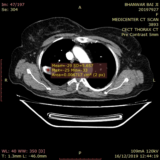
There is well defined lobulated solitary soft tissue lesion of size 17 x 35 x 26 mm in the right upper lobe. There are multiple areas of popcorn like calcification. Few areas also show fat attenuation(Upto-20 HU). The lesion shows mild enhancement on post contrast study. The lesion is extending upto the periphery of the lung and abutting the major fissure.
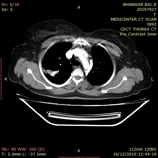
Few centrilobular nodules with ground glass opacities in right middle lobe.
The small area of fibro parenchymal thickening in the lingular segment of left upper lobe, suggest sequelae of old broncho-pulmonary infection.
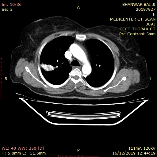
Rest of the lung appears normal with even distribution of the pulmonary arterial branches and that of the bronchial tree.
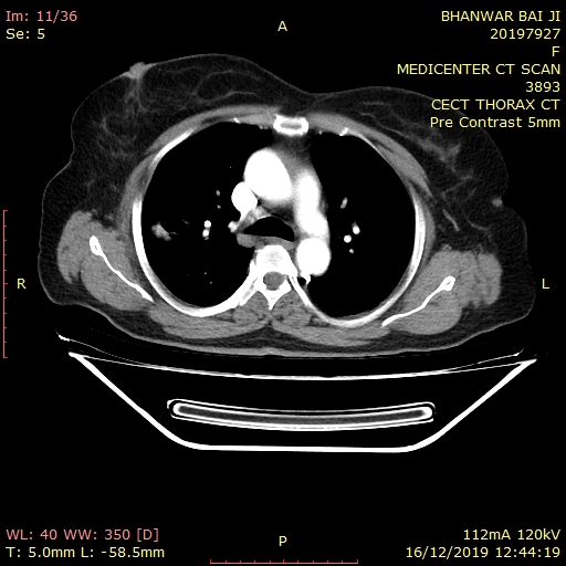
The anatomical configuration of the structures in the mediastinum and both hilar regions are normal.
There are subcentimeteric enlarged lymph nodes in pretracheal, right paratracheal & subcarinal region -?Reactive.
The arch of the aorta and the vessels arising from it are normal.
There is no evidence of pleural effusion seen.
Visualized abdomen shows no significant abnormality.
Bilateral adrenal glands are normal.
Visualized bones shows degenerative changes with diffuse idiopathic skeletal hyperostosis.
IMPRESSION: Above described CECT imaging findings are suggestive of
- Pulmonary hamartoma in right upper lobe.
- Few centrilobular nodules with ground glass opacities in right middle lobe, suggestive of infective etiology/bronchitis.
- Fibro parenchymal thickening in the lingular segment of left upper lobe, suggest sequelae of old broncho-pulmonary infection.