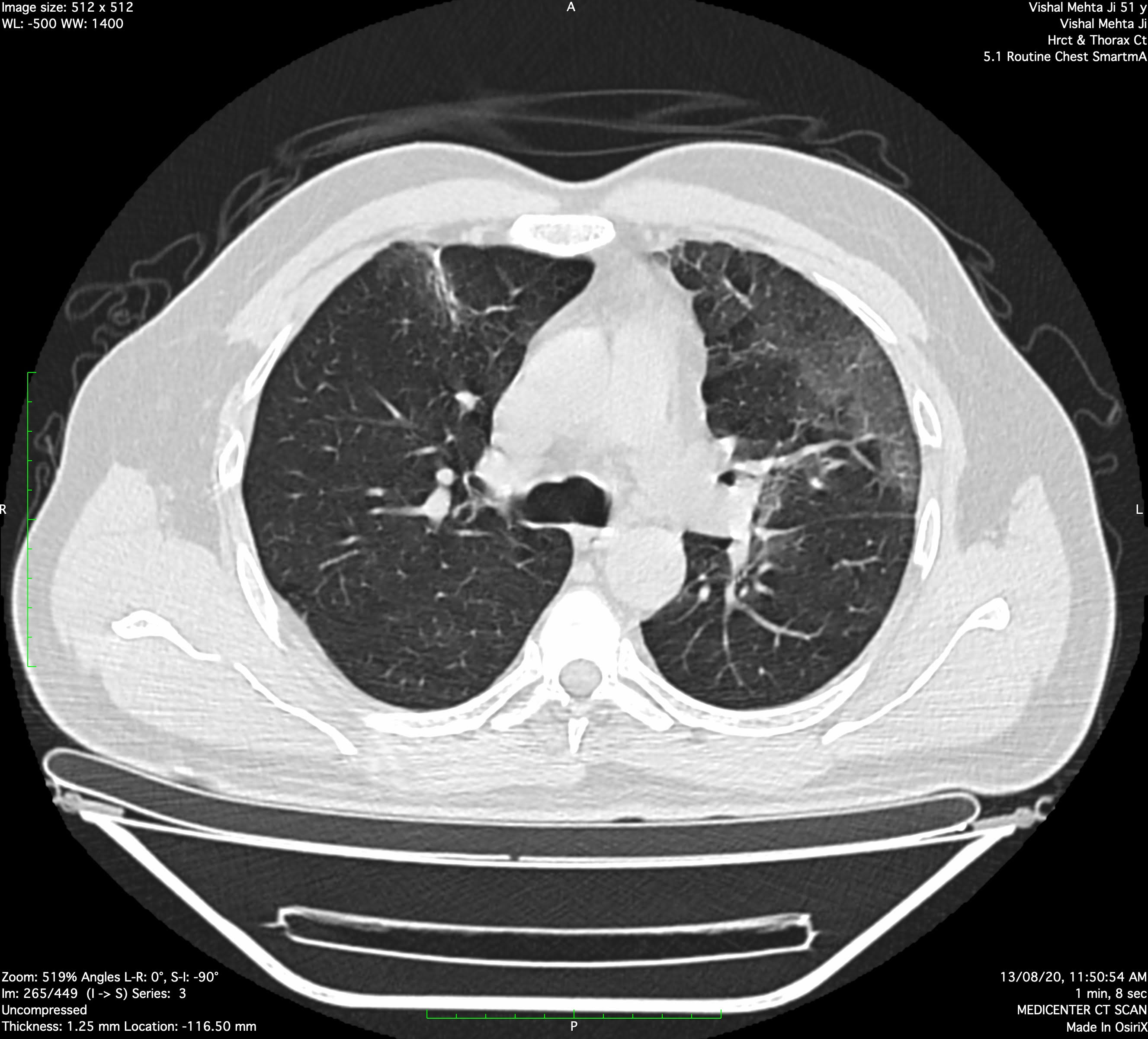
Multiple areas of subpleural and peri-broncho-vascular ground glass opacities with smooth interstitial septal thickening noted in medial and lateral segment of right middle lobe, superior basal segment of right lower lobe, apicoposterior, anterior & inferior lingular segment of left upper lobe, anterior basal, lateral basal and superior basal segment of left lower lobe.
Above findings suggest possibility of atypical pneumonia (intermediate to high suspicion of recent on going pendamic COVID-19 viral etiology). CORADS - 3/4.
Adv:- Clinico-pathological correlation
CT Severity:
Right upper lobe: 0-5 % (1 point)
Right middle lobe: 0-5 % (1 point)
Right lower lobe: 0 % (0 point)
Left upper lobe: 25-50 % (3 point)
Left lower lobe: 0 25 % (2 point)
Total CT severity scoring : 7 points (out of 25).
Read More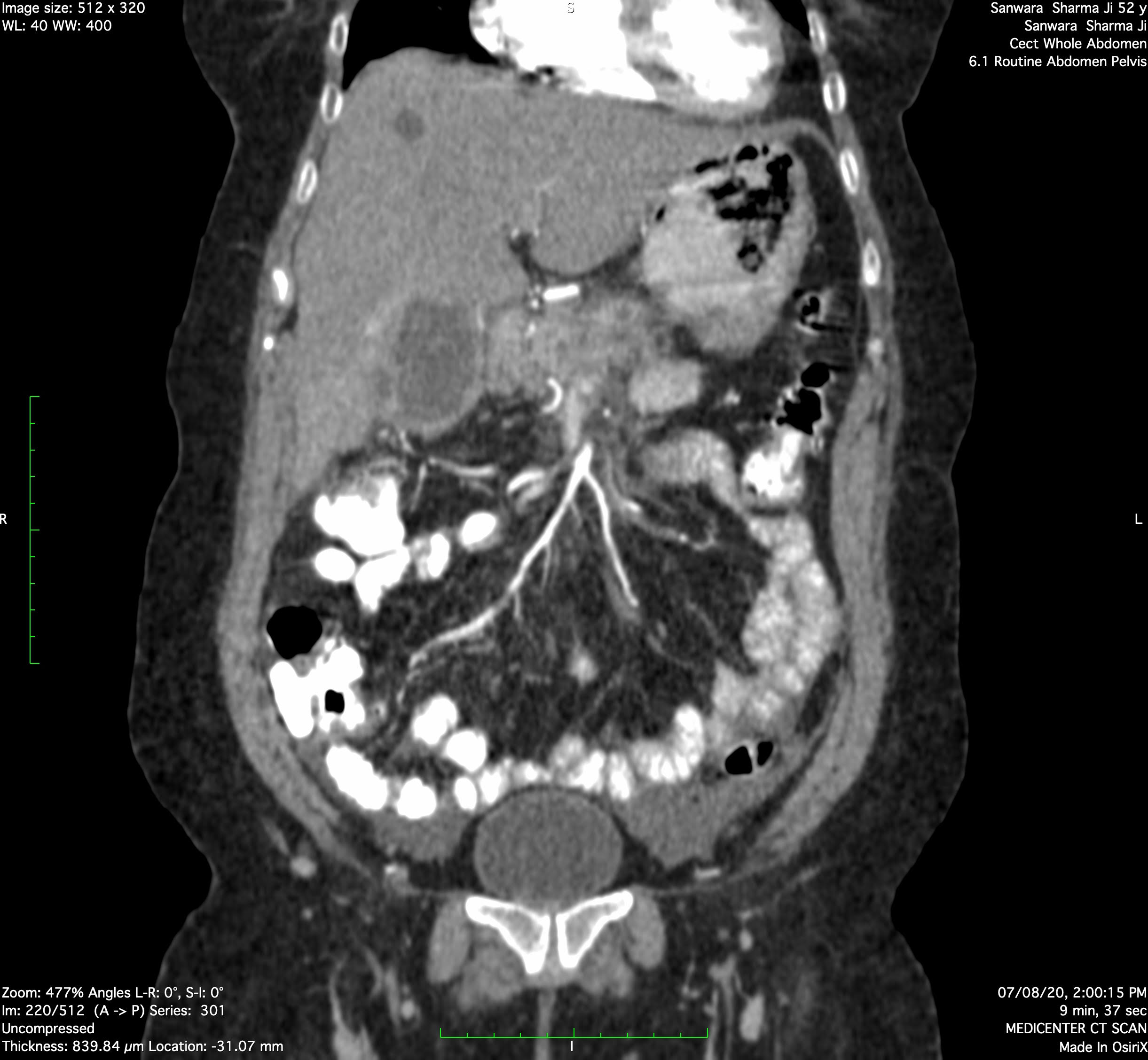
The gall bladder is overdistended with diffusely thickened & edematous gall bladder wall. Pericholecystic fluid & fat stranding noted. Findings suggestive of changes of cholecystitis.
Read More
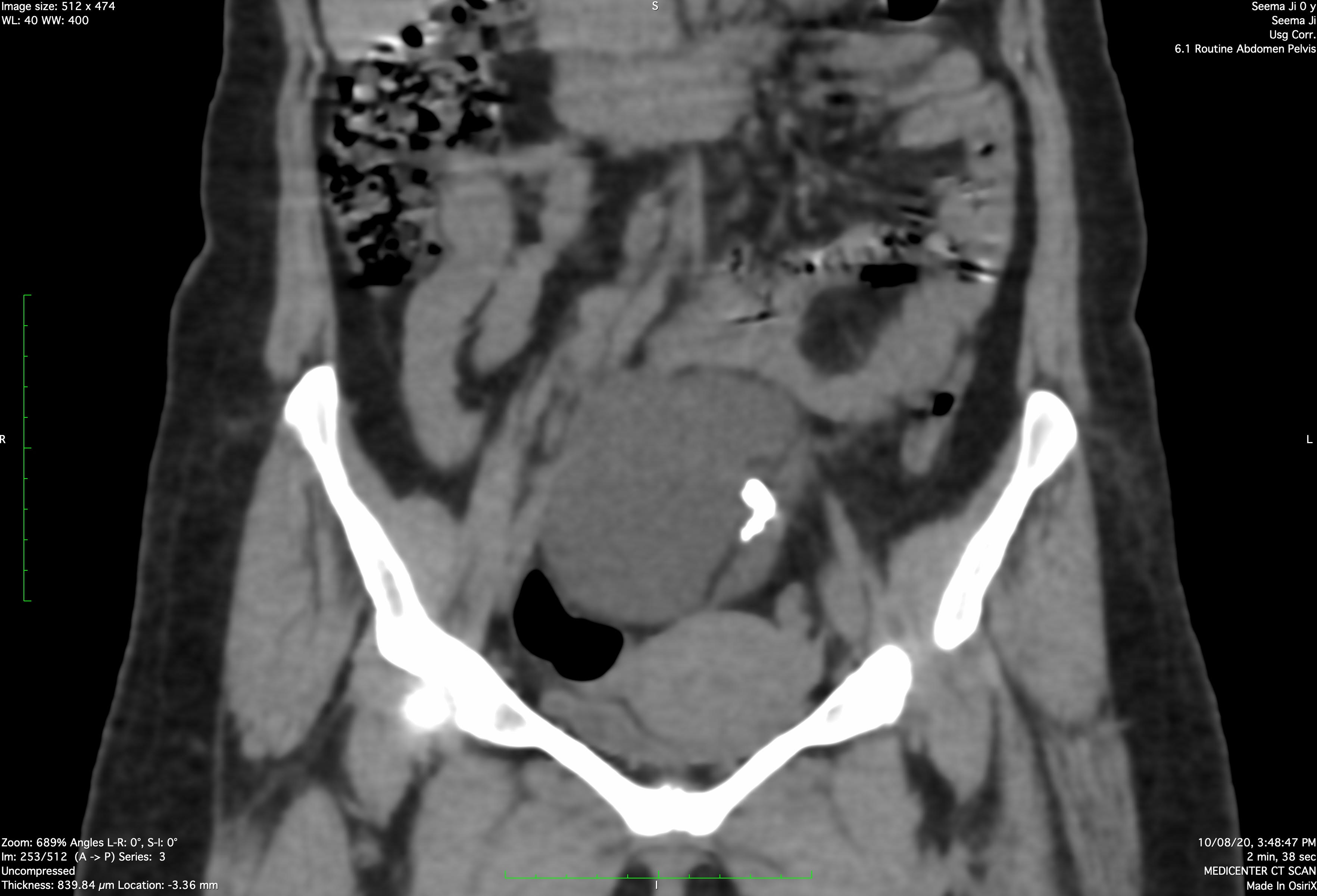
MRI OF PELVIS
Imaging Features:
Large altered signal intensity cystic lesion with internal septations and solid component (fat) noted in pelvis, possibly arising from right ovary causing compression and left lateral displacement of uterus. Lesion measures 9.3 x 11.3 x 10.8 cm. The lesion shows heterogeneous hypo to hyperintense signals on T1W & T2W images, few areas of suppression on STIR and blooming on FFE.
Suggest possiblity of right ovarian dermoid cyst
Read More
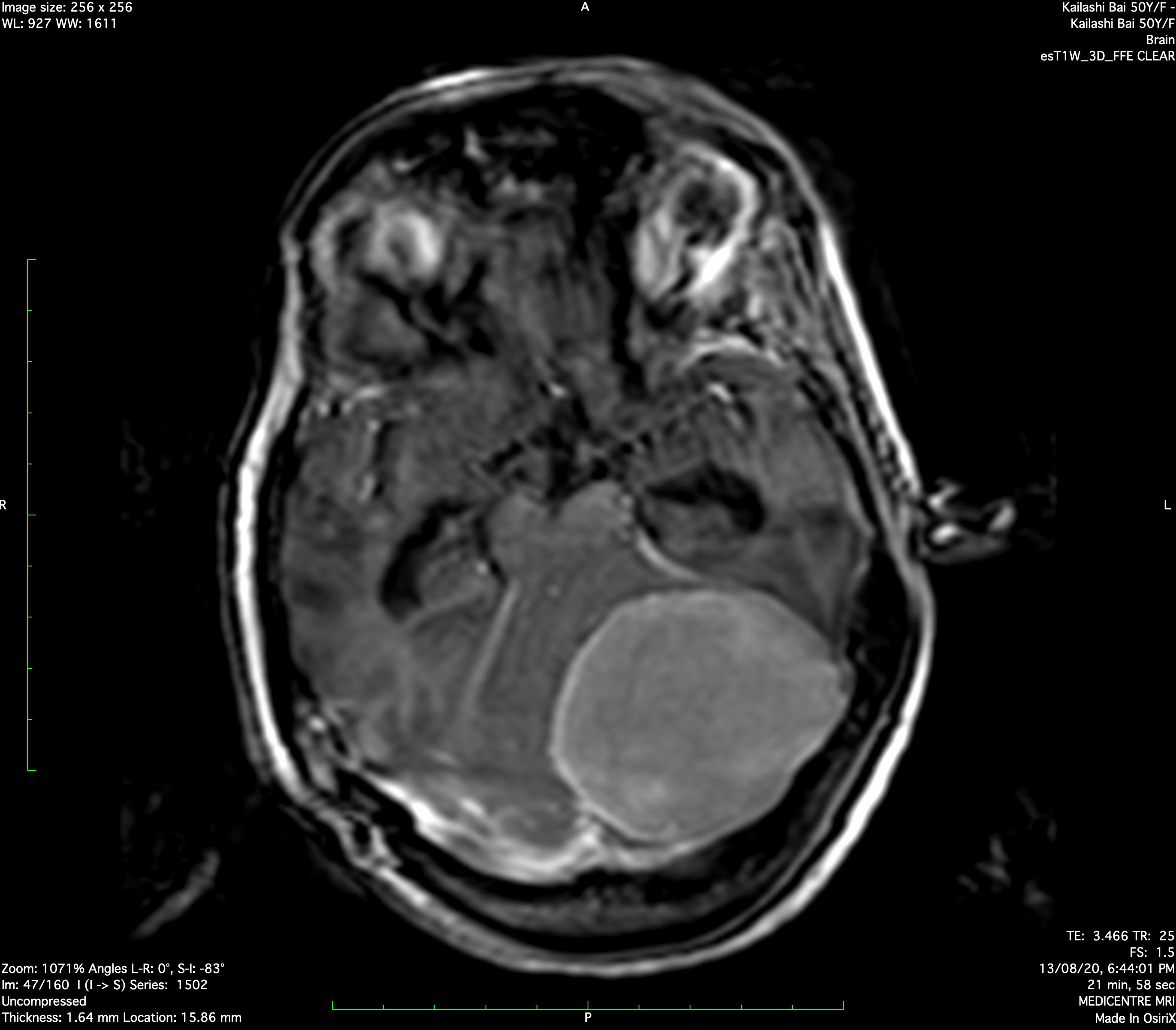
Imaging Features :
Well defined homogeneous enhancing extra axial altered signal intensity lesion noted along the posterior aspect of the left tentorium cerebelli. Lesion appears hyperintense on T2W images, iso to hypointense on T1W images. Lesion shows linear foci of FEE blooming likely calcification. Lesion measures approximately 5.2 x 5.4 x 5.2 cms (AP x ML x SI). Lesion causes mass effect in the form of effacement and compression over adjacent part of left cerebellar hemisphere, 4th ventricle and left half of brainstem.
Above finding suggest possibility of meningioma.
Read More
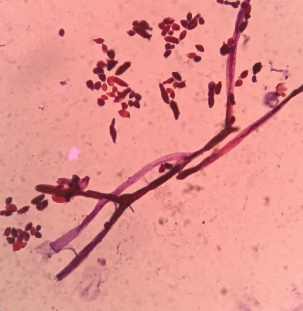
A 60 year male from rural area presented with complaints of multiple chronic discharging sinuses on his right foot . On biopsy , it was diagnosed as eumycotic myecetoma .
It’s fungal culture revealed septate hyphae with conidiogenesis .Colony characteristics and microscopic morphology was consistent with diagnosis of fungal etiology Exophiala jeanselme