C.T.SCANImaging Features :
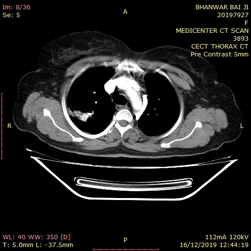
There is thick walled cavitatory lesion with spiculated borders in the apical segment of right upper lobe, measuring 50 x 48 x 51 mm. The lesion is causing stranding posterosuperiorly with infiltration & erosion of transverse process of D2, costovertebral joint and adjacent posterior 2nd rib & also erosion of the adjacent 3rd rib. Findings are suggestive of apical lung tumor (Pancost tumor). (Adv : HPE correlation)
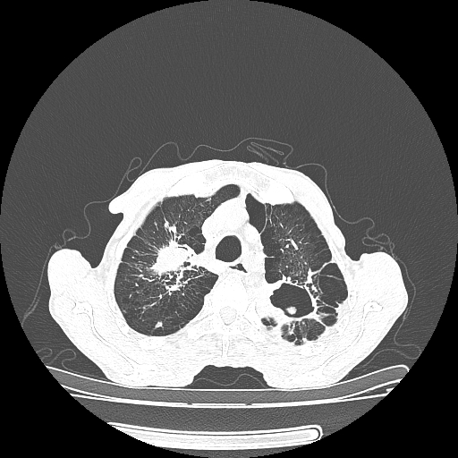
There are large fibrocavitatory parenchymal thickening with dystrophic calcification, bronchiectasis, subpleural consolidation with adjacent architectural distortion seen in the left upper lobe. Multiple centrilobular nodules few showing tree in bud pattern & calcification, bronchiectasis, peribronchial thickening, and interlobular interstitial thickening in both lungs. Finding are suggestive of post primary tuberculosis.
There is well defined oval shaped soft tissue density lesion of size 9 x 9 mm seen in the dependent part of the left upper lobe cavity, suggestive of aspergilloma .
Trachea mildly shifted towards right side
Centriacenar emphysematous changes are seen in both lungs.
There are few enlarged lymph nodes noted involving prevascular, pretracheal, bilateral paratracheal, AP window, precarinal & subcarinal regions, largest measures 15 x 10 mm in the subcarinal region, ? Granulomatous ? Metastatic
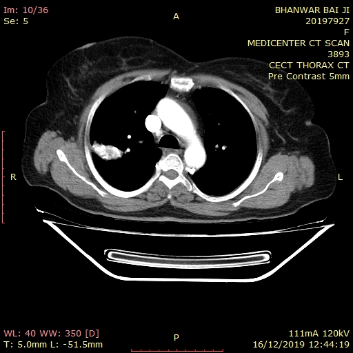
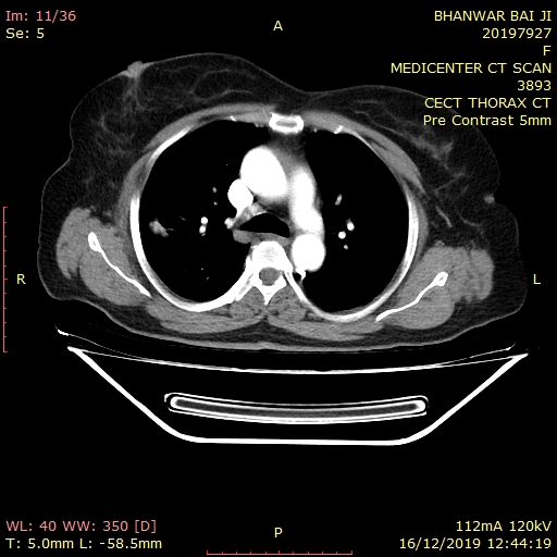
The arch of the aorta and the vessels arising from it are normal.There is no evidence of pleural effusion seen.
Note is made of sliding hiatus hernia.
Visualized abdomen shows bilateral simple renal cortical cysts.
Visualized bones show old united fracture in the left 11th & 12th ribs posteriorly.
Impression: Above described CT imaging features reveal
- Right apical lung tumor (Pancost tumor) as described above. (Adv : HPE correlation)
- Post primary tuberculosis with aspergilloma in left upper lobe cavity.
- Diffuse centriacenar emphysematous changes.
- Mediastinal lymphadenopathy ? Granulomatous ? Metastatic
- Sliding hiatus hernia.
- Bilateral simple renal cortical cysts.
- Old united fracture in the left 11th & 12th ribs posteriorly.