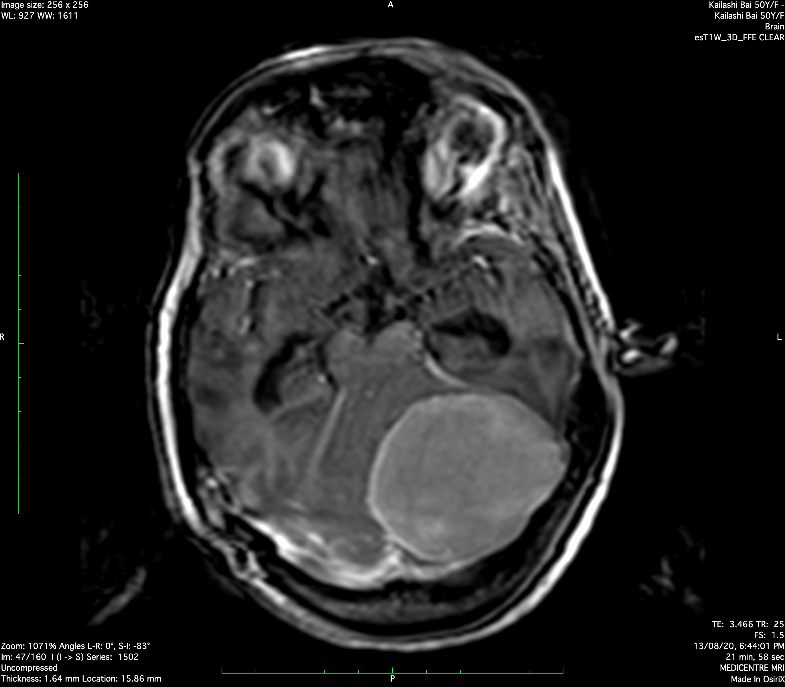


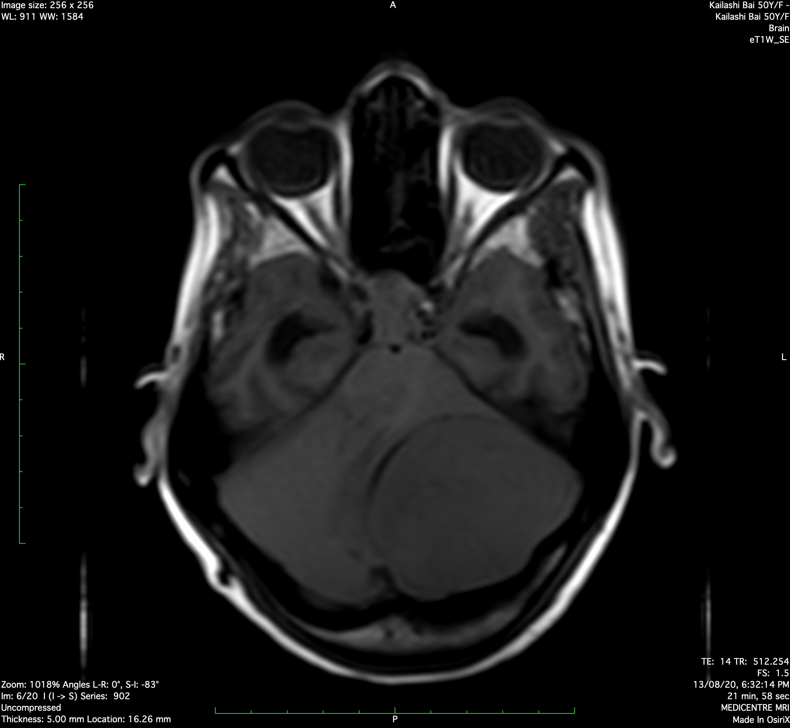
Imaging Features :
Well defined homogeneous enhancing extra axial altered signal intensity lesion noted along the posterior aspect of the left tentorium cerebelli. Lesion appears hyperintense on T2W images, iso to hypointense on T1W images. Lesion shows linear foci of FEE blooming likely calcification. Lesion measures approximately 5.2 x 5.4 x 5.2 cms (AP x ML x SI). Lesion causes mass effect in the form of effacement and compression over adjacent part of left cerebellar hemisphere, 4th ventricle and left half of brainstem.
Above finding suggest possibility of meningioma.