Ultrasound In Pregnancy
Ultrasound is a safe and non-invasive imaging method that utilizes sound waves to produce images of the growing fetus, placenta, and uterus. It allows physicians to monitor fetal growth, identify abnormalities, and estimate the due date for the baby. Throughout pregnancy, several ultrasounds may be scheduled at different stages for various purposes.
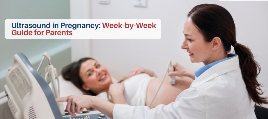
Here, provided for you, is a complete week-by-week guide for parents at each stage of ultrasound testing.
Importance Of Ultrasounds During Pregnancy
Ultrasounds in pregnancy are vital because:
- Confirms pregnancy and fetal and heartbeat.
- Checks the baby’s position, size, growth, and organ development.
- Detects multiple pregnancies like twins or triplets.
- Identifies potential abnormalities or complications.
- Estimates gestational age and due date.
- Monitors the placenta and amniotic fluid levels.
- Reveals your baby's genitals
- Guides procedures like chorionic villus sampling (CVS) or amniocentesis
Additional Note: In India, it is prohibited to determine the gender of the baby, under the Pre-Conception and Pre-Natal Diagnostic Techniques (PCPNDT) Act, 1994.
Week-by-Week Pregnancy Ultrasound Guide
At each stage of pregnancy, your baby will undergo a variety of unique developments. Ultrasound scans are used to monitor them all. Here is what to expect in each trimester:
1. First Trimester Ultrasound
-
Early Pregnancy (6-8 Weeks)
Often referred to as the viability scan or dating scan, this is the first ultrasound in pregnancy. It confirms that a gestational sac is present, identifies the presence or absence of a heartbeat, and confirms that the pregnancy is developing inside the uterus.
Key Highlights:
- Confirms intrauterine pregnancy.
- Detects multiple embryos.
- Estimates gestational age.
-
Nuchal Translucency Ultrasound (11-14 Weeks)
The baby nuchal translucency (NT) ultrasound is a scan that looks at the clear area at the back of a baby's neck. The primary purpose of the scan is to help doctors determine the likelihood of a baby having chromosomal abnormalities, such as Down syndrome.
Key Highlights:
- Assesses early fetal anatomy.
- Measures crown-rump length.
- Combined with a blood test for an accurate risk assessment.
2. Second Trimester Ultrasound
-
Anatomy Scan (18-22 Weeks)
The anatomy scan, which is also called the mid-pregnancy scan, is one of the most comprehensive ultrasounds. It looks at the baby’s organs, arms, legs, vertebrae, and brain to check if the baby has any growth abnormalities
Key Highlights:
- Evaluates fetal anatomy in detail.
- Checks placenta position and amniotic fluid.
- Determines fetal sex
Additional Note: Determining the sex of a baby is prohibited in India, under the Pre-Conception and Pre-Natal Diagnostic Techniques (PCPNDT) Act, 1994 3. Third Trimester Ultrasound
3. Third Trimester Ultrasound
-
Growth Scan (28-32 Weeks)
This ultrasound is an evaluation of the baby’s size: measurement of the head circumference, abdominal circumference, and the length of the femur to estimate the baby’s weight and growth trend. It helps ensure the baby is developing proportionately and identifies any signs of growth restriction or excessive growth.
Key Highlights:
- Measures fetal size, weight, and head circumference.
- Check the placenta function.
- Monitors amniotic fluid volume.
4. Doppler Ultrasound (32-36 Weeks)
A Doppler ultrasound checks blood flow in the umbilical cord, placenta, and baby’s brain. The procedure is used to uncover fetal distress or a restricted growth.
Key Highlights:
- Monitors oxygen and nutrient supply.
- Detects umbilical cord or placental issues.
- Ensures the baby’s well-being in high-risk pregnancies.
5. Late Pregnancy Scan (36-38 Weeks)
This scan will confirm the position of the baby as well as determining if it is in the head-down position safe for vaginal delivery. The ultrasound also evaluates the amount of amniotic fluid, the maturity and place of the placenta as well as the condition of the fetus in general.
Key Highlights:
- Check baby’s position
- Evaluates amniotic fluid levels.
- Determines placental location and maturity.
Conclusion
An ultrasound during pregnancy provides peace of mind to expecting parents and allows them to learn about their baby’s development. By knowing what each week-to-week scan means, you can stay informed, prepared, and connected with your growing baby.
Monitor Your Baby’s Health With Confidence!
Redcliffe Labs facilitates you with expert pregnancy week-by-week ultrasounds. They are safe, accurate, timely and reliable scans performed throughout every stage of pregnancy from certified professionals. Schedule your appointment now!
FAQs
1. How many ultrasounds are recommended during pregnancy?
Usually, 3-5 ultrasounds are recommended to be done in a normal pregnancy: an early pregnancy scan (6-8 weeks), a nuchal translucency scan (11-14 weeks), an anatomy scan (18-22 weeks), a growth scan (28-32 weeks) and a late pregnancy scan (36-38 weeks). Higher risk pregnancies may involve more.
2. Is it safe to have multiple ultrasounds while pregnant?
Yes. Ultrasound employs sound waves, not radiation, and is therefore safe for both mother and baby. Your provider will order the scans based on medical necessity to follow the growth of the baby. A regular ultrasound will assist in monitoring your baby’s development and well-being along the way.
3. How should I prepare for a pregnancy ultrasound?
Preparation for an ultrasound depends on the stage of pregnancy. During early scans you may be asked to drink water as a full bladder helps provide clear images. Additionally, you may need to expose your abdomen for the scan, so wear comfortable clothes. Always follow your doctor’s instructions, especially if specialized scans like Doppler ultrasound or NT scan are scheduled.
4. Can ultrasound detect all abnormalities in the baby?
No. Ultrasound does not detect all abnormalities in the baby. While ultrasounds can identify many structural or growth-related issues, they cannot detect all genetic or developmental conditions. Some problems may require additional tests like amniocentesis or chorionic villus sampling (CVS).
5. Will the ultrasound reveal the baby’s gender?
Yes. Ultrasound can help reveal the baby's gender in the second trimester. In India, knowing the sex of a child before birth is strictly prohibited. Imaging techniques like pregnancy ultrasounds primarily focus on assessing fetal health and development.
6. How long does each ultrasound usually take?
Most routine ultrasounds take between 15-30 minutes, depending on the type and stage of pregnancy. Specialized scans like Doppler studies may take slightly longer.
7. Can ultrasounds accurately predict the baby’s due date?
Early pregnancy scans, particularly the first-trimester scan, provide the most accurate estimates of gestational age and estimated delivery date. Early pregnancy scans also assess early signs of possible developmental issues, which can give you and your doctor valuable insights for a healthy pregnancy.”
8. Are there any risks associated with pregnancy ultrasounds?
When done correctly, Ultrasounds are safe, and there haven’t been any risks that have been proven. Nevertheless, unnecessary scans should be limited to avoid excessive exposure. Stick to the doctor’s advice regarding the time and the number of scans for the safety of both the mother and the baby.
9. Can ultrasound scans detect multiple pregnancies?
Yes. Early scans can identify twins, triplets, or higher-order multiples, allowing doctors to monitor each baby’s growth and development closely. Detecting this condition early allows for planning prenatal care and safe handling of complications.
Categories
LATEST from Blog
- CT Scan: What It Is, When You Need It & How It Helps Diagnose Diseases
- Prioritizing Your Well-being: A Guide to Preventive Health Checkups in Udaipur
- Automated Microbiology Testing in Udaipur with VITEK Technology
- How to Check Heart Blockage at Home & Cardiac Tests in Udaipur
- X-Ray for Lungs: Tuberculosis, Pneumonia In Udaipur